39 diagram for labelling microscope
Labeling the Parts of the Microscope | Microscope World Resources Labeling the Parts of the Microscope This activity has been designed for use in homes and schools. Each microscope layout (both blank and the version with answers) are available as PDF downloads. You can view a more in-depth review of each part of the microscope here. Download the Label the Parts of the Microscope PDF printable version here. Parts of the Microscope with Labeling (also Free Printouts) 5. Knobs (fine and coarse) By adjusting the knob, you can adjust the focus of the microscope. The majority of the microscope models today have the knobs mounted on the same part of the device. Image 5: The circled parts of the microscope are the fine and coarse adjustment knobs. Picture Source: bp.blogspot.com.
Microscope Types (with labeled diagrams) and Functions Simple microscope labeled diagram Simple microscope functions It is used in industrial applications like: Watchmakers to assemble watches Cloth industry to count the number of threads or fibers in a cloth Jewelers to examine the finer parts of jewelry Miniature artists to examine and build their work Also used to inspect finer details on products
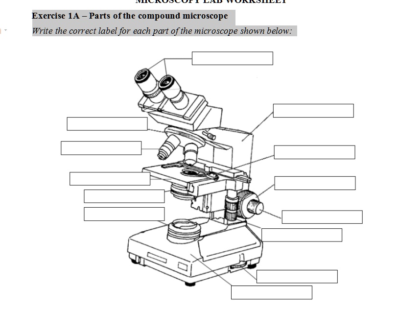
Diagram for labelling microscope
Deciphering the immunopeptidome in vivo reveals new tumour Verkko15.6.2022 · A newly developed genetically engineered mouse model enables the analysis of specific antigen presentation in vivo, providing insights into the tumour immunopeptidome and cancer progression. ZEISS LSM 980 with Airyscan 2 – Confocal Microscope with … VerkkoAiryscan 2 allows you to do more than any conventional LSM detector. Each of its 32 detector elements collects additional information, while all of them together gather even more light, yielding super-resolution quantitative results. Microscope Parts, Function, & Labeled Diagram - slidingmotion Microscope Parts Labeled Diagram The principle of the Microscope gives you an exact reason to use it. It works on the 3 principles. Magnification Resolving Power Numerical Aperture. Parts of Microscope Head Base Arm Eyepiece Lens Eyepiece Tube Objective Lenses Nose Piece Adjustment Knobs Stage Aperture Microscopic Illuminator Condenser Lens
Diagram for labelling microscope. Optical tweezers in single-molecule biophysics - Nature Verkko25.3.2021 · Optical tweezers have become the method of choice in single-molecule manipulation studies. In this Primer, we first review the physical principles of optical tweezers and the characteristics that ... Label the Microscope Diagram | Download Scientific Diagram - ResearchGate Label the Microscope Diagram Source publication +5 Laboratory Exercises in Microbiology: Discovering the Unseen World through Hands-on Investigation Book Full-text available Oct 2016 Joan... Compound Microscope Parts - Labeled Diagram and their Functions Labeled diagram of a compound microscope Major structural parts of a compound microscope Optical components of a compound microscope Eyepiece Eyepiece tube Objective lenses Nosepiece Specimen stage Coarse and fine focus knobs Rack stop Illuminator Condenser Abbe condenser Iris Diaphragm Condenser Focus Knob Summary An overview of microscopes Comparative genomic hybridization - Wikipedia VerkkoComparative genomic hybridization (CGH) is a molecular cytogenetic method for analysing copy number variations (CNVs) relative to ploidy level in the DNA of a test sample compared to a reference sample, without the need for culturing cells. The aim of this technique is to quickly and efficiently compare two genomic DNA samples arising …
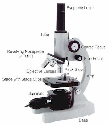
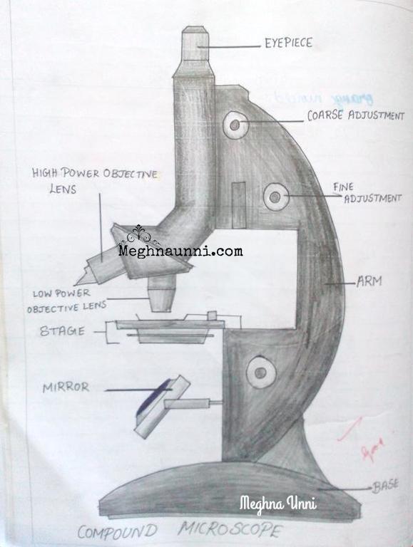
A Study of the Microscope and its Functions With a Labeled Diagram ... A Study of the Microscope and its Functions With a Labeled Diagram To better understand the structure and function of a microscope, we need to take a look at the labeled microscope diagrams of the compound and electron microscope. These diagrams clearly explain the functioning of the microscopes along with their respective parts. Labeling the Parts of the Microscope | Microscope World Resources Labeling the Parts of the Microscope This activity has been designed for use in homes and schools. Each microscope layout (both blank and the version with answers) are available as PDF downloads. You can view a more in-depth review of each part of the microscope here. Download the Label the Parts of the Microscope PDF printable version here. Microscope Labeling Diagram | Quizlet Coarse Focus Knob Moves the stage large distances to roughly focus the image. Fine Focus Knob Moves the stage tiny distances to slightly adjust and fine-tune the image focus. Arm Supports the body tube. Objective Lenses Focus and magnify light in differing amounts to view the specimen. Stage Clips Hold the slide in place on the stage. Nosepiece Labelled Diagram of Compound Microscope The below mentioned article provides a labelled diagram of compound microscope. Part # 1. The Stand: The stand is made up of a heavy foot which carries a curved inclinable limb or arm bearing the body tube. The foot is generally horse shoe-shaped structure (Fig. 2) which rests on table top or any other surface on which the microscope in kept.
Maternal inheritance of glucose intolerance via oocyte TET3 ... May 18, 2022 · e, The upper schematic diagram of mRNA micro-injection and oocyte maturation to investigate whether high glucose condition leads to the degradation of Tet3 transcript through its 3’-UTR. Microscope Parts and Functions Microscope Parts and Functions With Labeled Diagram and Functions How does a Compound Microscope Work?. Before exploring microscope parts and functions, you should probably understand that the compound light microscope is more complicated than just a microscope with more than one lens.. First, the purpose of a microscope is to magnify a small object or to magnify the fine details of a larger ... Electron microscope - Wikipedia VerkkoAn electron microscope is a microscope that uses a beam of accelerated electrons as a source of illumination. As the wavelength of an electron can be up to 100,000 times shorter than that of visible light photons, electron microscopes have a higher resolving power than light microscopes and can reveal the structure of smaller objects. A … Labelling a Microscope Diagram | Quizlet The function of the microscope stage is to allow for easy movement and manipulation of the slide. This will allow you to focus on the specimen in an accurate manner. What is the diaphragm? A diaphragm on a microscope is the piece that enables the user to adjust the amount of light that is focused under the specimen being observed. Light Source.
SOLVED: Exercise 1A Parts ofthe compound microscope Write the ...
European Commission VerkkoEuropean Commission - Policies, information and services. Select your language. български español čeština dansk Deutsch eesti ελληνικά English (Current language) français Gaeilge hrvatski italiano
Label a microscope - Teaching resources
Label the microscope — Science Learning Hub Label the microscope Interactive Add to collection Use this interactive to identify and label the main parts of a microscope. Drag and drop the text labels onto the microscope diagram. eye piece lens diaphragm or iris coarse focus adjustment stage base fine focus adjustment light source high-power objective Download Exercise Tweet

Microscope Labeling
Labelling A Microscope Teaching Resources | Teachers Pay Teachers Print & Go Notes - No Prep Needed!This resource includes student notes, a teacher key, and slides for teaching. It follows the Alberta Science 10 Unit C (Biology) Curriculum.Student Notes (7 pages) covers the following topics:microscope diagram - labelling parts and their functionstimeline of the development of microscopes & cell theorycalculations (magnification, field of view, actual size ...

Microscope label Diagram | Quizlet
Lifestyle | Daily Life | News | The Sydney Morning Herald VerkkoThe latest Lifestyle | Daily Life news, tips, opinion and advice from The Sydney Morning Herald covering life and relationships, beauty, fashion, health & wellbeing

label microscope diagram | Charts | Microscope, Diagram chart ...
Shared and distinct transcriptomic cell types across ... - Nature Verkko1.11.2018 · The neocortex contains a multitude of cell types that are segregated into layers and functionally distinct areas. To investigate the diversity of cell types across the mouse neocortex, here we ...

How to draw Microscope diagram for beginners - step by step
BCE Curriculum VerkkoReligious Knowledge and Deep Understanding. Familiarity with characters, events and messages from some key Old Testament stories, including Joseph (Genesis 37:1-36, 39:1-6, 41:15-44, 41:53-57, 42-46) and David (1 Samuel 17:1-49), is a means of connecting Scripture and real life.. View additional details about Literacy View …

Compound and Stereo- microscopes - Microscopes 4 Schools
Sperm Under Microscope with Labeled Diagram - AnatomyLearner The labelled diagram has already described all the structures of sperm in this article. Conclusion. So, this article provides the details structural features of sperm under the light microscope. All the labeled diagrams might help you identify the sperms from seminiferous tubules and epididymis of an animal.

13 - Microscope Parts - PowerPoint Worksheet | PDF | Glass ...
Simple Microscope - Parts, Functions, Diagram and Labelling Simple Microscope - Parts, Functions, Diagram and Labelling By Editorial Team March 7, 2022 A microscope is one of the commonly used equipment in a laboratory setting. A microscope is an optical instrument used to magnify an image of a tiny object; objects that are not visible to the human eyes. Table of Contents
Microscope Parts & Specifications | Microscope World Resources
Label the Light Microscope - Labelled diagram - Wordwall Label the Light Microscope. Share Share by Nquinn805. Show More. Like. Edit Content. Embed. More. Leaderboard. Show more Show less . This leaderboard is currently private. Click Share to make it public. This leaderboard has been disabled by the resource owner. This leaderboard is disabled as your options are different to the resource owner. ...

Compound Microscope Parts – Labeled Diagram and their ...
Simple Microscope - Diagram (Parts labelled), Principle, Formula and Uses A simple microscope consists of Optical parts Mechanical parts Labeled Diagram of simple microscope parts Optical parts The optical parts of a simple microscope include Lens Mirror Eyepiece Lens A simple microscope uses biconvex lens to magnify the image of a specimen under focus.
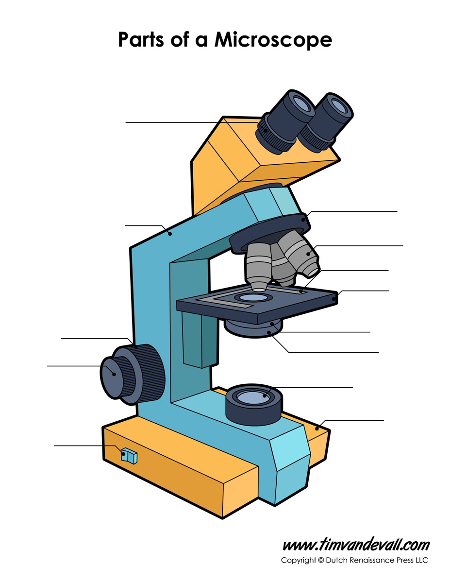
Microscope Diagram Labeled, Unlabeled and Blank | Parts of a ...
Fluorescence microscope - Wikipedia A fluorescence microscope is an optical microscope ... are of the epifluorescence design shown in the diagram. ... the main techniques are labelling with ...

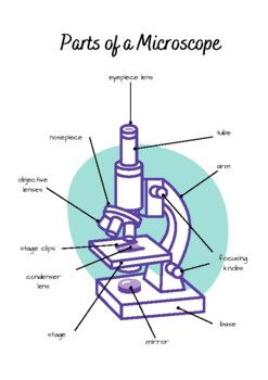
Parts of a microscope with functions and labeled diagram
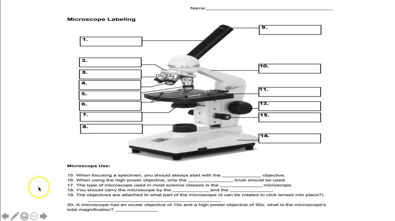
Microscope Labeling - The Biology Corner The labeling worksheet could be used as a quiz or as part of direct instruction where students label the microscope as you go over what each part is used for. The google slides shown below have the same microscope image with the labels for students to copy.
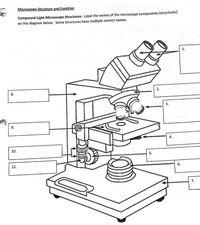
Answered: Microscope Structure and Function… | bartleby
Label Microscope Diagram - EnchantedLearning.com Using the terms listed below, label the microscope diagram. arm - this attaches the eyepiece and body tube to the base. base - this supports the microscope. body tube - the tube that supports the eyepiece. coarse focus adjustment - a knob that makes large adjustments to the focus. diaphragm - an adjustable opening under the stage, allowing ...
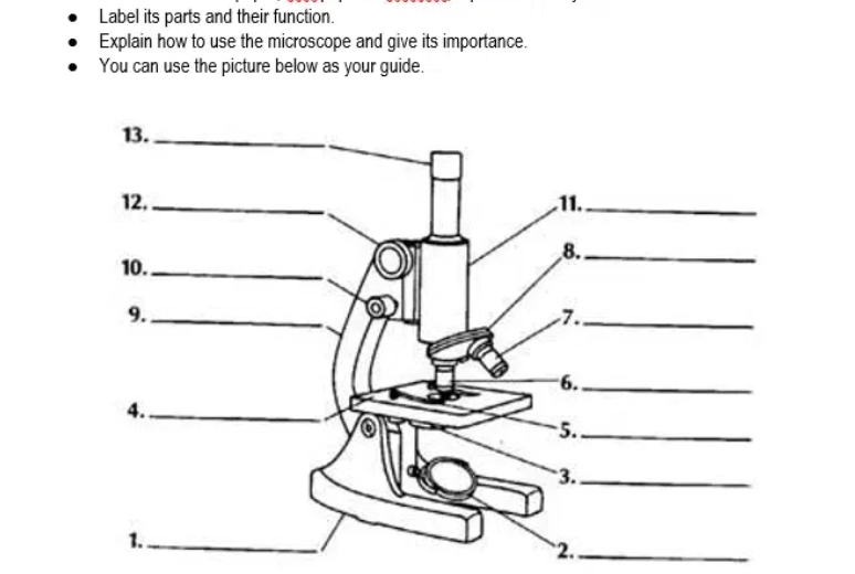
Answered: Label its parts and their function.… | bartleby
Parts of a microscope with functions and labeled diagram - Microbe Notes Parts of a microscope with functions and labeled diagram September 17, 2022 by Faith Mokobi Having been constructed in the 16th Century, Microscopes have revolutionalized science with their ability to magnify small objects such as microbial cells, producing images with definitive structures that are identifiable and characterizable.

Label the microscope — Science Learning Hub
Microscope Labeling Game - PurposeGames.com Microscope Labeling Game by sloanescience 2,105,364 plays 15 questions ~ 40 sec More 511 4.08 (you: not rated) Language English Tries Unlimited [?] Last Played November 23, 2022 - 03:36 PM There is a printable worksheet available for download here so you can take the quiz with pen and paper. Remaining 0 Correct 0 Wrong 0 Press play! 0% 0:00.0
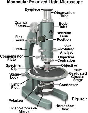
Molecular Expressions Microscopy Primer: Specialized ...
Microscope labeled diagram - SlideShare Microscope labeled diagram Oct. 30, 2013 • 6 likes • 28,252 views Download Now Download to read offline Pisgah High School Follow Advertisement Recommended Microscope Basics Mrs. Henley 3.4k views • 7 slides Parts and Functions of the Compound Microscope IsaganiDioneda 3.3k views • 43 slides SCIENCE7: The Microscope Christian Adriano-ursabia

File:Labelledmicroscope.gif - Wikimedia Commons
Microscope Labeling - The Biology Corner Microscope Labeling Microscope Labeling Microscope Use: 15. When focusing a specimen, you should always start with the _____________ objective. 16. When using the high power objective, only the _______________ knob should be used. 17. The type of microscope used in most science classes is the ______________ microscope. 18.

This is a common compound microscope. Label its parts from A ...
Microscope, Microscope Parts, Labeled Diagram, and Functions Microscope, Microscope Parts, Labeled Diagram, and Functions What is Microscope? A microscope is a laboratory instrument used to examine objects that are too small to be seen by the naked eye. It is derived from Ancient Greek words and composed of mikrós, "small" and skopeîn,"to look" or "see".

Microscope Labelling Review Diagram | Quizlet
Label Microscope Diagram - EnchantedLearning.com Label Microscope Diagram Using the terms listed below, label the microscope diagram. Inventions and Inventors arm - this attaches the eyepiece and body tube to the base. base - this supports the microscope. body tube - the tube that supports the eyepiece. coarse focus adjustment - a knob that makes large adjustments to the focus.
Microscope - Teaching resources
Whole-animal connectomes of both Caenorhabditis elegans sexes Verkko3.7.2019 · Quantitative connectivity matrices (or connectomes) for both adult sexes of the nematode Caenorhabditis elegans are presented that encompass all connections from sensory input to end-organ output ...
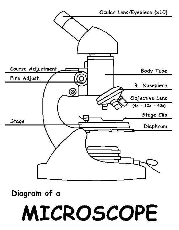
Diagram of a Microscope by ScienceDoodles on DeviantArt
Microscope Parts, Function, & Labeled Diagram - slidingmotion Microscope Parts Labeled Diagram The principle of the Microscope gives you an exact reason to use it. It works on the 3 principles. Magnification Resolving Power Numerical Aperture. Parts of Microscope Head Base Arm Eyepiece Lens Eyepiece Tube Objective Lenses Nose Piece Adjustment Knobs Stage Aperture Microscopic Illuminator Condenser Lens

Label the Microscope Diagram | Download Scientific Diagram
ZEISS LSM 980 with Airyscan 2 – Confocal Microscope with … VerkkoAiryscan 2 allows you to do more than any conventional LSM detector. Each of its 32 detector elements collects additional information, while all of them together gather even more light, yielding super-resolution quantitative results.

Can someone can send me diagram of this compound microscope ...
Deciphering the immunopeptidome in vivo reveals new tumour Verkko15.6.2022 · A newly developed genetically engineered mouse model enables the analysis of specific antigen presentation in vivo, providing insights into the tumour immunopeptidome and cancer progression.
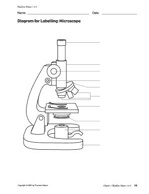
Fillable Online Blackline Master 12-3 - Richmond School ...

Label Microscope Diagram - EnchantedLearning.com

Simple Microscope - Diagram (Parts labelled), Principle ...
Microscope labelling - Teaching resources

Diagram of a Compound Microscope

Microscope Components - Science Quiz
Labelling A Microscope Teaching Resources | Teachers Pay Teachers
Biology : Compound Microscope Diagram for Class 8 ...
Microscope Labeling

Compound Microscope Parts

Carl Zeiss Microscopy Optical microscope Worksheet Diagram ...
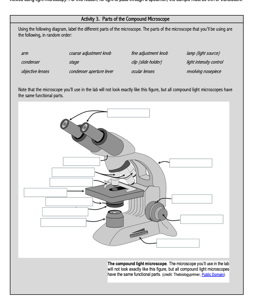
Solved Activity 3. Parts of the Compound Microscope Using ...

Compound Microscope- Definition, Labeled Diagram, Principle ...

Microscope Labeling Activity - SMART Board Activity - Interactive Review

Draw a well labelled diagram of a microscope. - Brainly.in

A Study of the Microscope and its Functions With a Labeled ...


Komentar
Posting Komentar